Modris Baum's Photo Gallery
MDL-K6GBaryte BaSO4 , Calcite CaCO3

Field of View: 3.1 mm
Largest Crystal Size: 1.0 mm
Found Oct 1992.
This is a comparison of MW UV fluorescence and visible light images.
This specimen is from the same find as [https://www.mindat.org/photo-1254387.html], which see for details. The MW response of the baryte in the current image looks more bluish than shown under this link, but I think that’s mostly because those crystals have thing coatings of Fe oxides which makes them look yellowish.
Both the indigo-violet and red responses in the background are probably from calcite. (I didn’t test the red stuff for reaction to HCl – there is very little of it on the specimen. But other calcite on this and other specimens from this find that I did test had a wide variety of responses ranging from red to indigo, with some specimens – including this one – hosting tiny cavities with very tiny bright yellow fluorescing crystals.)
2F4-KMFScapolite, Calcite CaCO3 , Graphite C , Muscovite KAl2(AlSi3O10)(OH)2


Field of View: 6.7 mm
Found 5/19/02.
This is a comparison of MW UV fluoresce (top), visible light, and SW UV fluorescence (bottom) images. With the 5 W LED flashlights that I use, the MW response is brighter than the SW response, but both “scapolite” responses are very bright. See below for details.
In both UV images, the yellowish glow is from “scapolite” (not analyzed). In these images the MW and SW colors look almost identical, but to the naked eye, the MW response seems more “saturated”. The other fluorescent mineral in these images is calcite – strong magenta-pink under MW; weak reddish under SW.
The main scapolite crystal is well-terminated and nearly complete. The three calcite crystals are all partial rhombs – well formed on top, but contacted on the bottom. The only other mineral visible in these images is graphite – the dark crystals seen nearly edge on at upper right.
These crystals are part of a very large (23 x 10 x 6 cm) specimen, one side of which consists almost entirely of scapolite, about 40% of which fluoresces bright yellow (as shown here) and the rest of which fluoresces slightly weaker and paler yellowish. Here and there on this side, there are small calcite crystals such as the one shown here as well as small graphite crystals. The other side of the specimen consists of large cleavages of calcite which fluoresce strong magenta to violet-blue under MW and very weak magenta under SW. These layers are separated by a layer of non-fluorescent brownish-green micaceous stuff.
Update April 23 2024: When I posted these images, I had forgotten that material from this specimen was analyzed EDS) in early 2022. The results suggested that the “scapolite” was more likely marialite than meionite and that the “mica” was muscovite var phengite. Some of this “mica” occurs as single crystals with rounded cross-sections embedded in the calcite. Many of these embedded crystals are nearly transparent and yellowish – but still not fluorescent. Another specimen, found the same day, has muscovite crystals as large as 2.6 cm. (See [https://www.mindat.org/photo-445175.html]. These look a lot like phlogopite, but they too have no UV response (MW or SW). (I will post the analytical data as time permits.)
Numerous small graphite crystals, some with interesting growth patterns are also embedded in the calcite.
The “scapolite” crystals shown here grew on one side of the top layer. There are a few larger crystals that are also well-terminated and nearly complete, but they are too large to photograph using a scope. Most of the scapolite in the top layer is so intergrown that individual crystals can’t be discerned.
The UV images were made with the flashlights positioned ca 5-6 cm from the specimen using objective magnification 2X on my scope with ISO 800 on my camera. Each frame for the (stacked) SW image required an exposure of 2.5 sec. Each MW frame required only 0.6 sec. Hand-held, the MW response looks about as bright as depicted with the flashlight held more than 1 m from the specimen. For similar brightness under SW, the flashlight has to be with 20-25 cm of the specimen.
Update April 24th 2024: I have now posted the analytical results from 2022 as child images. Both the EDS and Raman scans for the “scapolite” strongly suggest marialite dominant over meionite. But since this is not WDS data, I have posted it under “scapolite”. The scans for the “muscovite” are somewhat more ambiguous but still fairly convincing.
RFW-3CCCalcite CaCO3

Field of View: 6.55 mm
Found Oct 1992.
This is a comparison of MW UV fluorescence and visible light images. The calcite has no discernible SW response.
This specimen is from the same find as [https://www.mindat.org/photo-1367916.html] –which see for more info.
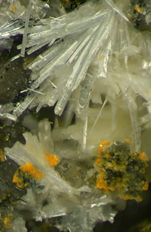
QT9-T6VCalcite CaCO3 , Pyrite FeS2

Field of View: 3.65 mm
This is a comparison of MW UV fluorescence and visible light images. (There is no SW response from the calcite.)
The MW response shown is somewhat similar to that of the “peanut” calcite from the “Buckwheat Dolomite” shown under [https://www.mindat.org/photo-1267045.html]. But this specimen is from the Lime Crest quarry – which is not exactly known for micros. More specifically, they come from a very tough, but slightly vuggy, boulder of tightly intergrown pyrite (see [https://www.mindat.org/photo-433989.html]) which also hosted the only euhedral baryte crystals that I ever found here. (The baryte is also fluorescent. See [https://www.mindat.org/photo-1254387.html].)
Note that there is some pyrite at upper right in the visible light image. Some of it looks tabular to me, but in other places, the pyrite clearly looks like intergrown cubes. (It has not been analyzed.)
The specimen also has a vein of violet-blue fluorescent calcite (MW only) - typical of the Franklin Marble. In places, the violet-blue grades to bright red – but not to the magenta-red shown here.
In addition to the cavities hosting euhedral calcite and baryte, there are some very small cavities (<= 5 mm) which have not only tiny calcite crystals, but also other glassy crystals that fluoresce very bright yellow. At this point, I don’t know what they are.
The MW response of the calcite crystals looks about as bright as depicted with a 5 W LED flashlight held as far as 25-30 cm from the specimen (in a room with some daylight). In making the photo image, I used objective magnification 4X, which “dilutes” the strength of the UV response significantly. Theefore I had use 8 sec exposures with ISO 800 when the flashlight was positioned ca 6 cm from the specimen.
RDC-0XPAllactite Mn2+7(AsO4)2(OH)8 , Roxbyite Cu9S5


Field of View: 6.7 mm
Via Nick Zipco (1993).
This is a comparison of visible light and MW UV fluorescence images. There is no SW image because bright green fluorescing spots (presumably willemite) all over the cavity side of the specimen make it impossible to see the SW fluorescent response of anything else. (Much of the embedded willemite on this specimen fluoresces very bright green under both SW and MW UV, but the willemite in the allactite cavity seems to have a rather modest MW response, so it does not obscure the other responses.)
In the visible light image, the reddish-brown crystals (mostly on the left) are allactite. In the MW image these crystals glow deep “burgundy red”. But the MW image is a bit of a fraud. It is actually almost impossible to see the response of the allactite when the specimen is hand-held or under a scope. (See below for details.) However, that doesn’t mean that the response isn’t “real”. In fact, in the photo image, the allactite response looks almost as bright as the yellowish/pinkish/greenish response of the (unidentified) spheroids. This response is easily seen with the naked eye with the MW flashlight held as far as 10-12 cm from the specimen – even with some daylight in the room. But to see the allactite response at all, the flashlight has to be almost touching the specimen and the room has to be completely dark. Why the difference? Perhaps it has to do with the sensitivity of my camera’s sensor to various wavelengths or the with the white balance settings that I used - I’m not sure.
Under visible light, the yellowish fluorescent spheroids look like grayish aggregates of tiny crystals. Under very high magnification, I can almost make out the shapes of these crystals – but not quite. A small sample – which broke off easily – dunked in HCl had no reaction at all. I have no idea what this stuff is.
Also in the visible light image, the bluish stuff at bottom center and right is roxbyite (possibly on chamosite or vermiculite). (EDS & XRD verified – see [https://www.mindat.org/photo-1247556.html].) In the MW image, the roxbyite looks as if it might have a weak violet-blue response, but that is almost certainly due to reflected visible light that go through the flashlight’s not too effective filter. There are also numerous thin, encrusted, “rods” scattered all over the allactite cavity that may have a weak bluish MW response. When dunked in HCl, the encrustation quickly vanishes, but I don’t see any fizz. The residue is a thin, round, colorless rod that appears to have no UV response at all. This is another UK – at least to me.
Not visible in these images are some bright blue and magenta-pink fluorescent blobs. Under a scope, these appear to be fine grained – no apparent cleavage. But they fizz vigorously and vanish completely in HCl – just like calcite.
There are many other aggregates of allactite crystals on this specimen. The ones shown in these images have the “brightest” UV response. Other crystals (see the child comparison pairs) have even weaker responses, so it is not surprising that fluorescent allactite has not been previously noted. As already mentioned, the response is only visible under MW and it doesn’t exactly “leap out” at you. However, the exposure settings need to make this image (6 sec per frame using ISO 800) were not particularly extreme. Even taking Photoshop post-processing, which roughly doubled the effective exposure time, into account, the required exposure wasn’t unusual. I have photographed many minerals that were easily naked eye visible which required similar or even longer exposures. (Magnification under a scope “dilutes” the UV responses, so sometimes the responses can be seen when a specimen is hand-held but hardly at all under the scope.)
8X6-W3GZincite ZnO , Willemite Zn2SiO4

Field of View: 1.75 mm
Thanks to Ralph Thomas.
The top MW UV mage shows a fleet of flying saucers in formation coming in for a landing on my microscope stage. (Who knew aliens were so tiny?) The bottom visible light image purports to show hemimorphic zincite pyramids. You decide.
RM5-X1YPascoite Group, Metamunirite NaVO3 , Bluestreakite K4Mg2(V4+2V5+8O28)·14H2O

Field of View: 1.35 mm
Thanks to Travis Olds.
This is a high magnification (objective 7X) stereo pair attempting to show the morphology of the tiny orange “pascoite group” crystals. Unfortunately, with my scope, “high magnification isn’t the same as “high resolution”. If you can fuse the images, that will help de-clutter the image and also make it look a bit sharper. But if you can’t, that’s OK.
Based on what I can see on other parts of the specimen and these images, I could be convinced that the “pascoite group” Is magnesiopascoite. But the fact is that I know nothing about this locality or about vanadium minerals, so “caveat emptor”. On the bottom right, the “pascoite group” appears to be mixed with - or even replacing – something else.
The much larger whitish crystals are probably metamurinite. They fluoresce bright crimson/magenta-red under MW UV and less bright red-orange under SW UV. The dark “blebs” are minor bluestreakite. See [https://www.mindat.org/photo-1366983.html] for details regarding the IDs and the strengths of the UV responses.
294-8RABluestreakite K4Mg2(V4+2V5+8O28)·14H2O , Metamunirite NaVO3 , Pascoite Group

Field of View: 2.25 mm
Thanks to Travis Olds.
This is a stereo pair. Things will look clearer and sharper if you can fuse the images, but if you can’t do 3D, that’s OK.
The dark crystal in the center is bluestreakite. It isn’t very big, but it may be of interest because it seems to be a single crystal and because it is from the TL. The white sprays are probably metamurinite. The crystals appear to be fairly robust for the species. Moreover, they fluoresce fairly bright magenta-red under MW UV (see the child MW/VL comparison pair) and less bright red-orange under SW UV. See [https://www.mindat.org/photo-1366983.html] where you will also find a discussion of the IDS and the strengths of the UV responses. The orange crystals are probably tiny “pascoite group”. Under very high magnification, a few appear to be well formed, but still too small for a definitive visual ID – not that I know anything about vanadium minerals anyway.
FGV-09RMetamunirite NaVO3 , Bluestreakite K4Mg2(V4+2V5+8O28)·14H2O , Pascoite Group

Field of View: 4.55 mm
Thanks to Travis Olds.
This is a comparison of SW UV fluorescence, visible light, and MW UV fluorescence images. With the 5 W LED flashlights that I use, the actual MW response is stronger than the SW response. See below for details.
In both UV images, the reddish glow is from metamurinite. As far as I can tell, these responses have not been previously reported, which is somewhat surprising because both responses are easily visible even with some daylight in the room. (Note: The metamurinite ID is visual by me, based on images of similar material posted by Alex Earl, Dan Polhemus, and others. See [https://www.mindat.org/photo-1067582.html] and [https://www.mindat.org/photo-943960.html] resp. )
Also visible in the UV images is a mineral that appears to have a weak grayish response under SW and a somewhat stronger violet-blue response under MW. Both of these responses are marginal and might possibly be “artifacts”. The MW response, in particular, might just be from reflection of violet-blue light that got through the MW flashlights visible light filter. The response looks “real” to me, but I’m not sure. (The violet streaks seen on some of the metamurinite are clearly due to reflect light. They move around if the flashlight is positioned differently.) In any case, real fluorescence or not, I have no idea what this stuff is. Under a scope using visible light, I see faintly violet-gray “blobs” with no distinct outlines. I couldn’t find anything on the Blue Streak Mine species list that looks similar.
In the visible light image, the bluestreakite is the very dark stuff; the “pascoite group” is orange. The latter is another visual ID by me. (Travis’ label only lists the bluestreakite.) Neither the bluestreakite nor the “pascoite” fluoresces. The “pascoite” crystals are very tiny, but some appear to be well-formed. Under very high magnification, a few of them look like they might be magnesiopascoite, but I don’t really know anything about this mine or about vanadium minerals in general, so consider that just a wild guess.
I have also posted a child MW/VL/SW comparison triplet of images showing another part of the specimen.
With my LED flashlights positioned ca 5-6 cm from the specimen, using objective magnification on my scope, and ISO 800 on my camera, each frame for the (stacked) SW image required an exposure of 13 sec. Each MW frame required only 2 sec. (Note: Exposure times vary greatly depending on how accurately one aims the flashlights. Also, the relatively long exposure time required for SW images is probably due, at least in part, to the fact that SW unit uses a Hoya filter. That filter is much more effective at blocking unwanted violet & blue light from the LED, but it probably also blocks more of the UV.)
When hand-held, the MW response is about as bright as depicted with the flashlight as far as 20-25 cm from the specimen – even in a room with dim daylight. For the SW response to look as bright, the flashlight has to be within 3-4 cm of the specimen.
1W1-Y4AEpididymite Na2Be2Si6O15·H2O , Calcite CaCO3 , Catapleiite Na2Zr(Si3O9)·2H2O

Field of View: 7.0 mm
Found Sept 1996.
This is a comparison of visible light and SW (middle) & MW (bottom) UV fluorescence images. It also a new parent photo, posted to show the very unusual whitish SW response of the epididymite. The violet MW response is also unusual, but in this image it looks “foggy” and indistinct because the brighter magenta response of calcite is lighting up the edges of the epididymite “snowflake”. Before I accidentally bumped it, I had another specimen with a “snowflake” that grew perpendicular to the matrix so that it was possible to see the MW response quite clearly against a dark background. But I never got to photograph it ... (All of the previous images have been retained as child photo. Note that the epididymite “snowflake has also lost a “propeller” since it was last photographed!)
Also visible in all the images are small (pseudo-)hexagonal crystals of catapleiite. These fluoresce yellowish-green on the edges only.
In general, epididymite and eudidymite from MSH do not fluoresce. But there are a few exceptions. Bright white fluorescing eudidymite is shown under [https://www.mindat.org/photo-1262635.html]. Bright green fluorescing epididymite is shown under [https://www.mindat.org/photo-1264766.html] and [https://www.mindat.org/photo-1290103.html].
With the flashlights positioned ca 5-6 cm from the specimen, using objective magnification 2X on my scope and ISO 800 on my camera, each frame for the (stacked) SW image required an exposure of 13 sec. Each MW frame required only 1.3 sec. When hand-held, the MW response looks about as bright as depicted with the flashlight as far as 30-35 cm from the specimen. To see a SW response roughly as bright, the flashlight has to be within 2-3 cm of the specimen.


