Home PageAbout MindatThe Mindat ManualHistory of MindatCopyright StatusWho We AreContact UsAdvertise on Mindat
Donate to MindatCorporate SponsorshipSponsor a PageSponsored PagesMindat AdvertisersAdvertise on Mindat
Learning CenterWhat is a mineral?The most common minerals on earthInformation for EducatorsMindat ArticlesThe ElementsThe Rock H. Currier Digital LibraryGeologic Time
Minerals by PropertiesMinerals by ChemistryAdvanced Locality SearchRandom MineralRandom LocalitySearch by minIDLocalities Near MeSearch ArticlesSearch GlossaryMore Search Options
The Mindat ManualAdd a New PhotoRate PhotosLocality Edit ReportCoordinate Completion ReportAdd Glossary Item
Mining CompaniesStatisticsUsersMineral MuseumsClubs & OrganizationsMineral Shows & EventsThe Mindat DirectoryDevice SettingsThe Mineral Quiz
Photo SearchPhoto GalleriesSearch by ColorNew Photos TodayNew Photos YesterdayMembers' Photo GalleriesPast Photo of the Day GalleryPhotography
╳Discussions
💬 Home🔎 Search📅 LatestGroups
EducationOpen discussion area.Fakes & FraudsOpen discussion area.Field CollectingOpen discussion area.FossilsOpen discussion area.Gems and GemologyOpen discussion area.GeneralOpen discussion area.How to ContributeOpen discussion area.Identity HelpOpen discussion area.Improving Mindat.orgOpen discussion area.LocalitiesOpen discussion area.Lost and Stolen SpecimensOpen discussion area.MarketplaceOpen discussion area.MeteoritesOpen discussion area.Mindat ProductsOpen discussion area.Mineral ExchangesOpen discussion area.Mineral PhotographyOpen discussion area.Mineral ShowsOpen discussion area.Mineralogical ClassificationOpen discussion area.Mineralogy CourseOpen discussion area.MineralsOpen discussion area.Minerals and MuseumsOpen discussion area.PhotosOpen discussion area.Techniques for CollectorsOpen discussion area.The Rock H. Currier Digital LibraryOpen discussion area.UV MineralsOpen discussion area.Recent Images in Discussions


EducationChert/chalcedony thin sections

30th Sep 2018 17:36 UTCNorman King 🌟 Expert

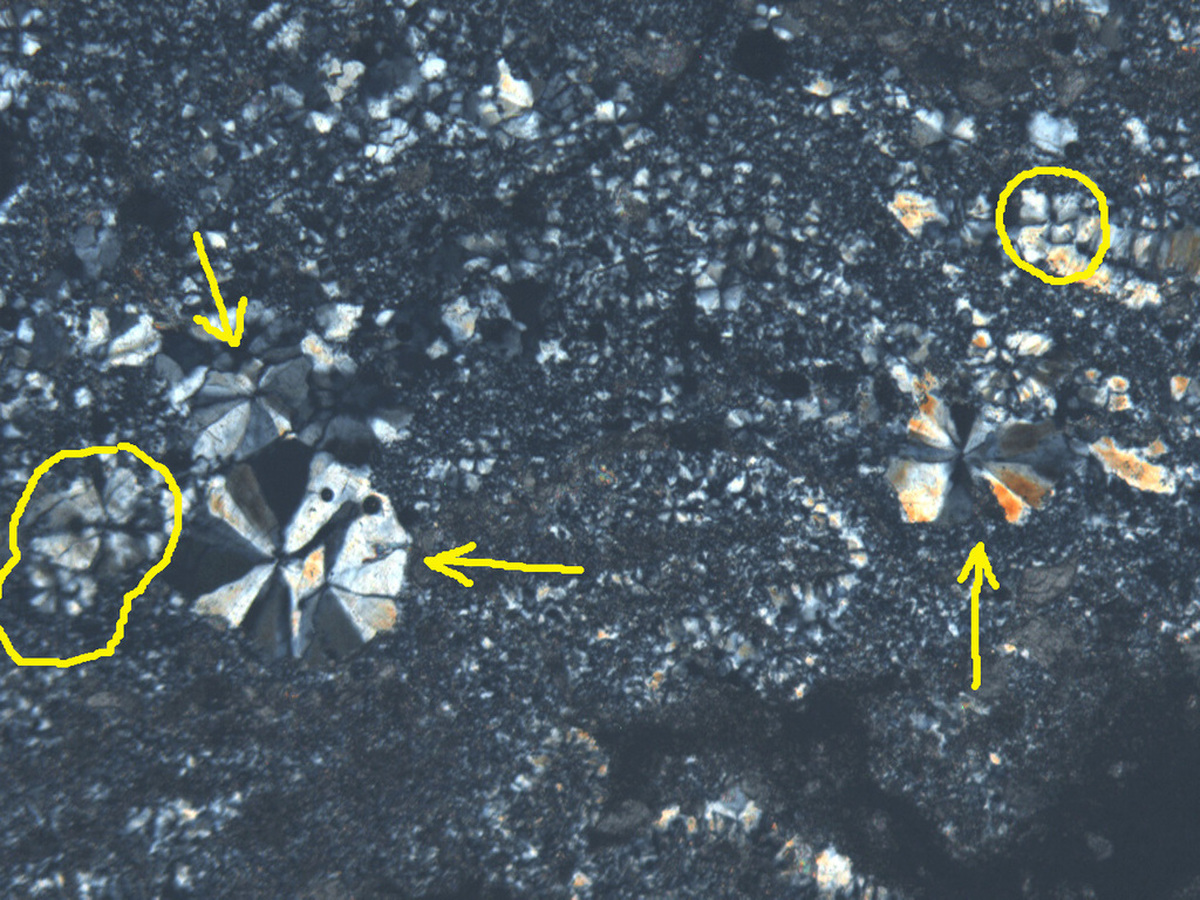
The second photomicrograph is at about 15% greater magnification. Fields of view are 3.0 mm (above ) and 2.6 mm (below) respectively. Several spheres that are about twice as large as those shown in the first photomicrograph are shown here (yellow arrows). Their crosses are less developed than those in the first photomicrograph. The component crystals here appear solid, so the quartz is more like that in chert than chalcedony. I was surprised to see similar extinction crosses (is that the right term?). I see only a few that are just like those in the first photomicrograph (some are circled here). Is this the same phenomenon in these two types of artifacts?

30th Sep 2018 18:30 UTCDon Saathoff Expert

1st Oct 2018 00:01 UTCBen Grguric Expert
1st Oct 2018 01:34 UTCNorman King 🌟 Expert

Thanks for your input. I’ll check length-slow vs. length-fast tomorrow.
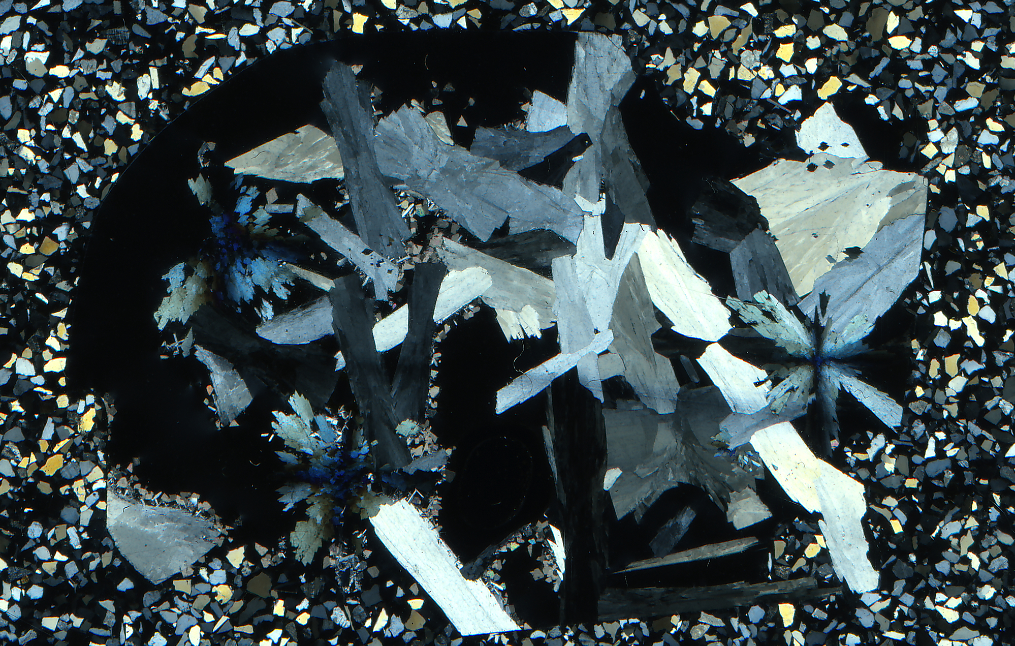
These two photomicrographs are from one thin section from the McNamara Formation (Belt Supergroup) near Missoula, Montana. The sample was sent to me by Daniel Bennett, who had started the "silicified mudstone?" thread a few months ago about identifying chert by its "hardness."
Both of these photos show the transition from fibrous chalcedony to what many people might call chert. Sorry the one is so dark–I’m trying to avoid optical “bloom.” Actually, the matrix really is dark when using crossed polarizers. I’ll have to experiment with my photographic system to see if I can do better. Note that in the first view–a closeup of the transition zone (FOV is only 1.9 mm)–the more cherty-looking blocky grains seem to have quadrate form, as if they are breaking up along the extinction crosses. Of course that doesn’t make any sense if those crosses are merely optical artifacts that exist only within my microscope! Are these quadrate shapes an illusion aided by undulatory extinction? The extinction crosses and quadrate shapes are less obvious as the microscope stage is rotated. It may be that the four “subcrystals” of one cross “system” are amplified optically, but only when in this position and every 90 degrees from there around 360 degrees. They are least obvious, or not visible at all, when the microscope stage is at a 45 degree angle from these positions.
The view below (FOV is 5.4 mm) provides a better feel for the context. Portions of the larger field of small cherty crystals still seem to have a quadrate tendency. That’s a more subjective judgment, however, especially when considering the larger field.
transition from chalcedony to chert
1st Oct 2018 02:20 UTCRichard Gibson 🌟
http://edafologia.ugr.es/optmine/xplconos/futallw.htm
The others may be the same, just in more complex crystals.
2 cents
cheers
dick gibson
1st Oct 2018 02:28 UTCBen Grguric Expert
1st Oct 2018 22:28 UTCNorman King 🌟 Expert
Incidentally, I had a thin section handy of chert ("flint") from chalk of the Paris Basin (France), and tested several recrystallized fossil fragments in it, finding some microspheres (I do not know what animal/plant group those came from) that are length-slow. Most of the fossil debris I tested there is length-fast.
Richard, I wonder about the uniaxial interference figures. I don't see any color bands, or halos, in any of these. Should I, if that's what they are?
6th Oct 2018 20:18 UTCNorman King 🌟 Expert

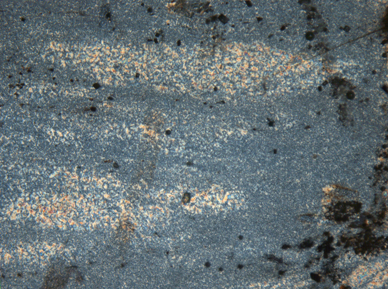
The dark figures in circular crystallites I showed in a thin section of the Gunflint chert from Blende Lake, Ontario do not change position or orientation as the microscope stage is rotated. Moreover, all of those crystallites have the same pattern, even though optic axes of individual crystals (fibers) presumably are varied with respect to the thin section orientation. Apparently the most important condition for appearance of these pseudo-interference figures (the term I will use for this phenomenon) is a habit of fibrous crystals in radial arrangement. Those crystals (fibers) positioned parallel to microscope polarization directions (due east-west and due north-south) do not allow the passage of polarized light. Crystal fibers must be oblique to directions of polarization to allow polarized light through. The pseudo-interference lines (the so-called Maltese cross) are commonly bent or otherwise distorted. Most pseudo-interference figures in the Blende Lake thin section are bent.
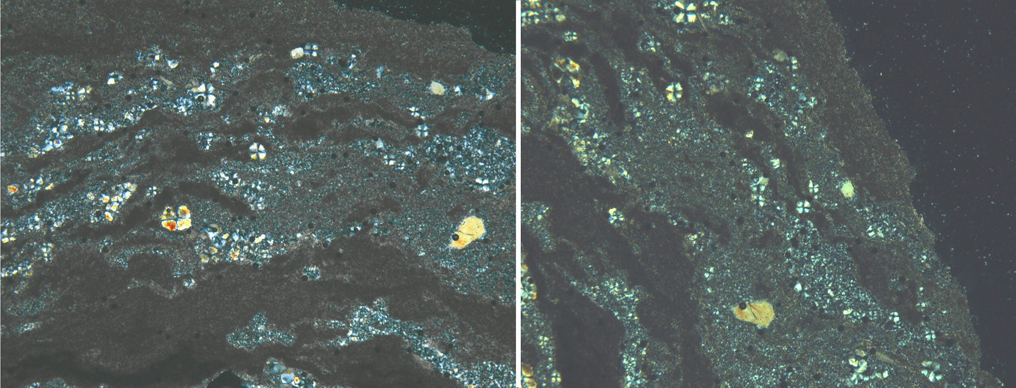
The thin section from Missoula County, Montana (see first additional thin section view below) shows generally equidimensional quartz microcrystals tightly packed together. Local groups of larger chalcedony/chert crystals seemingly form tetrads of four squarish crystals separated by the pseudo-interference lines. Some of those lines are strongly distorted. Strong undulatory extinction may be at least partially responsible for that. Commonly two adjacent, relatively distinct members of a tetrad actually consist of partially merged tetrads, making some appear to have six members (sextets). In places, trains of such merged tetrads make two-dimensional, ladder-like series (see second additional thin section view below). Note that many of the pseudo-interference figures in the second new thin section are highly distorted. Some "tetrads" seem to have just three members, instead of four. Others look like there may be two that are at least partially superimposed in nearly the same place. I attribute these issues to reflect distortion of the crystallites (i.e., the fibers are no longer straight, if they ever were) or periodic growth of crystallites in the same area, perhaps allowed by continued or periodic supply of fluids.
For all of these, the pseudo-interference lines typically become disrupted as the microscope stage is rotated, but not in a systematic way. This makes it hard to keep track of individual members of the tetrads and even whole tetrads during stage rotation. Rotating the stage exactly 90 degrees (or 180 or 270) usually yields a “picture” that can be readily matched with the original view.
Comments?
Reference: West, I. (2018). Geology of the Wessex Coast (part of Jurassic Coast, Dorset and East Devon World Heritage Site. http://www.southampton.ac.uk/~imw/Portland-Isle-Geological-Introduction.htm (Version 15th June, 2018)

Chalcedony Blende Lake

6th Oct 2018 23:24 UTCRalph S Bottrill 🌟 Manager
I think Ben and yourself have got it, these are pseudo-interference figures commonly seen in any radial-fibrous anisotropic minerals under cross-polarised light due to the alignment of the optic axes with the polarisers. I often feel such silica microspherules are replaced microfossils but am open to suggestions on that. I tend to think we should call the recrystallised silica cherty quartz rather than chert, as the latter is a rock name, and can comprise mostly chalcedony, but maybe that will start an argument?
6th Oct 2018 23:50 UTCFrank K. Mazdab 🌟 Manager
Interference figures are an optical effect caused by strongly converging light passing through a single crystal. The interesting "cross" effect seen here (and in most transparent minerals occurring as radial or spherical clusters) is simply the result of extinction of a portion of an aggregate of fine-grained mineral fibers oriented parallel to the microscope's N-S and E-W polarizers. The same thing can be seen in zeolites and other minerals that take on this macroscopic texture. In balls of cavansite, the mineral's extreme dispersion frames the cross in electric blue and brown (although because the crystals are coarser the cross is not as sharp). Next time I'm on back the microscope (Monday?), I'll update this post with a photo, or even take a video and link to it here, since as we can see from the previous posted photos, the effect is quite attractive and appears to inspire conversation.
I don't think the effect requires a special name, but if you want to give it one, I'd suggest "radial aggregate extinction".
7th Oct 2018 03:30 UTCNorman King 🌟 Expert
Ralph, Good to hear from you again. The way you said what you said impressed me more this time than the last time we knocked chert and chalcedony around several years ago. I think I see at least undulatory extinction in most or all cherts for which I have cut thin sections–25 or so. There may or may not be some local concentrations of fibrous material in them. But, as I understand this now, chert is the rock name, and chert consists mostly of the mineral chalcedony, by definition. In other words, it appears that “chert” cannot be applied to a single microcrystal of quartz. The aggregate is chert. Do I have that right?
I am concerned that the same issue will arise with the name chalcedony, however. I don’t think people are used to calling a rock consisting largely of botryoidal (fibrous!) chalcedony “chert.” I think they want that stuff to be chalcedony, whether used as a rock name or mineral name. So then we would have the term (chalcedony) referring to both a mineral and a rock.
A different but somehow similar situation applies to salt vs. rock salt, and gypsum vs. rock gypsum. No one seems to have problems with those. But quartz vs. rock quartz? No way!--It’s quartz and chert! All the while, no one is clamoring for different names for hematite–one for megacrystals and one for fibrous botryoidal hematite (or malachite, or several zeolites, etc.).
P.S., Ralph: I think the replaced microfossils in chert would have (or had at one time) distinct spherical enclosures, with chalcedony crystallizing within those and then replacing the “shell.” There should be clues to that–different crystal size, orientation, color, inclusions, or something else–maybe isotopes.
9th Oct 2018 02:32 UTCFrank K. Mazdab 🌟 Manager
24th Oct 2018 01:02 UTCNorman King 🌟 Expert
Enough of that. I took these photomicrographs of a thin section of chert, not expecting any complications to arise. I labeled the first one as at zero degrees (or I should say that I turned the microscope stage to zero degrees and took a photo there. Then I turned the stage 45 degrees and took another photo. Wow, what a difference! The chalcedony shows more interference colors. Areas that were various shades of gray in the first photo became lit up mainly in yellow and red in the second photo. Yellow and red are the first colors to show, then purple and blue, going from first to second order interference colors.
First photo at zero degrees on the microscope stage.

chalcedony TS from McNamara-2
Photo at the 45 degree position. Yes, this is the same field of view!
When I turned the microscope stage another 45 degrees, it returned to what I saw at the zero spot. That was 90 degrees from the first photo. Then, after another 45 degrees (135 degrees altogether) it looked like the view at 45 degrees. And so on for 360 degrees around. BTW, above I showed the second photo (the microscope stage rotated 45 degrees) in the whole rectangular visual field. Below is the same field with rotation to the opposite direction by 45 degrees using Photoshop that returned all landmarks to the same as in the original photo for ease of comparison.

chalcedony TS from McNamara-3
Second photo rotated in opposite direction as microscope stage using Photoshop to return all landmarks to same positions as in original photo.
It seems that all the chalcedony crystals are in similar alignment. My guess is that chalcedony crystals or crystal groupings in this sample of chert (the rock!) have become preferentially aligned in a kind of foliation that is visible using cross-polarized light in the petrographic microscope. This sample is from the Mesoproterozoic McNamara Formation of the Belt Supergroup near Missoula, Montana. So it had plenty of time to have suffered directed stress, perhaps either from the same stress field that caused thrusting in this area, or simply from burial metamorphism.
What about you petrographers out there? Any thoughts about this effect?
P.S. Frank, Thanks for uploading the additional photo showing cavansite with radial aggregate extinction and stilbite, to say nothing of the hundreds of rock photomicrographs you uploaded and showed us where to find!
27th Oct 2018 00:29 UTCNorman King 🌟 Expert
Radial crystallites of chalcedony produce extinction crosses in cross-polarized light that I referred to earlier (this thread, October 06, 2018 08:18PM) as “pseudo-interference figures.” Frank Mazdab, who knows more about thin section petrography than I, called this phenomenon “radial aggregate extinction.” Ideally, those extinction crosses depend on orientation of the polarizer and analyzer in the petrographic microscope, and should be visible in radial crystallites of other minerals besides chalcedony, as Frank pointed out. The arms of the crosses should always be north-south and east-west, meeting at a central point (“origin”), no matter how the rock is oriented. Below is the composite of two photos of Gunflint chert from Blende Lake, Ontario, Canada that illustrate this principle. The crosses are north-south and east-west in both photos even though the two views are oriented about 55 degrees from each other. So, these two photos show chalcedony crystallites doing the right thing! FOV for left photo is 3.0 mm.
Initially in this study I took seven photomicrographs of the same field of view in chert from McNamara, Missoula County, Montana, each photo being rotated 15 degrees on the microscope stage from the previous one. After all of that photography I determined that just the zero-degree, the 45-degree, and 90-degree views are sufficient to make my point, so the composite photomicrograph below includes only those three photos. Too many photos that differ by too little, in a field of view where you can easily have difficulty in following the "behavior" of each crystallite is a lot of work for those of us who have little background in examination of high-magnification thin sections. To review the vocabulary, the thin sections here show the rock chert, composed of crystallites of presumably fibrous chalcedony (a variety of quartz). The left-to-right field of view in the first (left-hand) photo is 1.7 mm. The red “plus” sign in the middle photo shows how the pseudo-interference crosses should be oriented in all three views. Note the dark branching feature in each view that you can use to verify that this is indeed the same field of view, and you can use it as a reference for orienting yourself with respect to crystallites in each view. The view at zero degrees (left photo) and 90 degrees (right photo) are nearly identical. You just have to rotate your head 90 degrees clockwise to see the same view in the right photo as in the left photo. Crystallites are in the same positions and pseudo-interference crosses are similar. Granted the crosses are crude, showing curved lines, and some are disjointed at their origin. In contrast, the middle view at 45 degrees doesn’t match the other two very well at all. If you tilt you head 45 degrees clockwise, the same crystallites will be difficult to identify, and some of the pseudo-interference crosses are impossible to locate. A few of the larger crystallites are recognizable in the middle view even if they are smaller and the crosses are not quite oriented due north-south and east-west, some are curved or bent, and the origin may be disjointed. Note that the dark marker “branch” is right where it should be in all three views–just rotated.
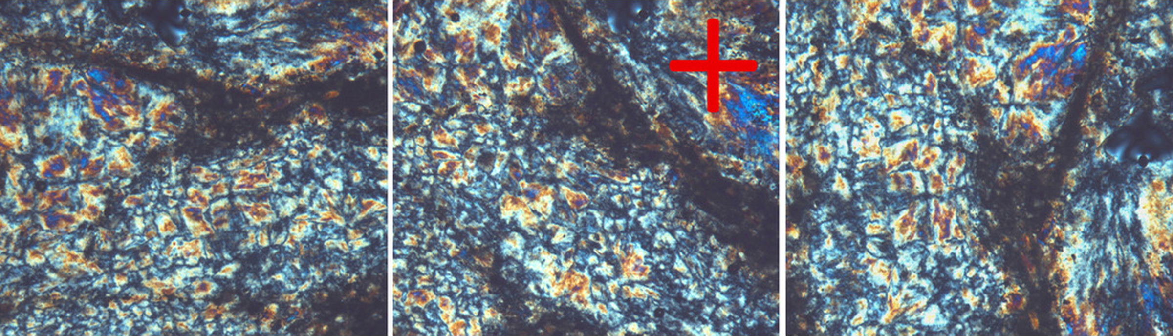
Composite photo of chalcedony crystallites
Maybe the crystallites have been smooshed by some kind of deformation. As I suggested in my posting of October 24, 2018 01:02AM in this thread, metamorphism or other regional stresses may have caused a grain (foliation?) to develop or have otherwise played havoc with the ideal geometry.
Or, maybe they aren’t really made of radial fibrous crystals. In fact, many of the larger crystallites clearly consist of four smaller crystals, making a unit resembling the Windows trademark of four panes in a window. Where some of those are right next to each other they tend to run together so that the individual smaller four panes seem to belong to two different larger “windows” at the same time. I haven’t seen reference to such quadrate “windows” before. I suggest they form by breakup of silky bundles of fibrous chalcedony crystals in the process of forming more equidimensional crystals that would be more stable in terms of boundary energy.
The next composite of two photos below, also from McNamara MT, include a zero-degree view (left photo) in which there are many quadrate window-like units having more or less north-south and east-west radial arms, but some of those are bent and others do not originate evenly at the center of the crosses. The 45-degree view (right photo) has very few north-south, east-west crosses, and the ones that can be seen are poorly developed. Details in the 45-degree view lack clarity and are so unlike what is visible in the zero-degree view that I used numbers to mark seven corresponding spots in both views that are opaque impurity spots not tied to any chalcedony crystals or crystallites. I linked those spots with lines, the series of which altogether lie at an angle of approximately 45 degrees from each other in the two views to help people oriented themselves. FOV for each photo in the pair is 1.7 mm
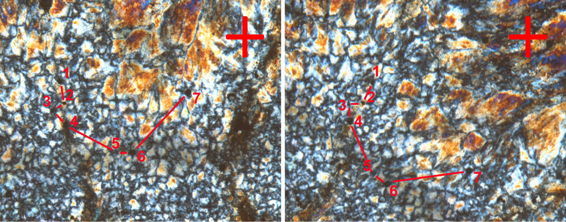
Composite photo of chalcedony crystallites-2
So, to be on the record, I have illustrated these phenomena and suggested what is going on.
Anybody else have any ideas?
28th Oct 2018 14:51 UTCNorman King 🌟 Expert

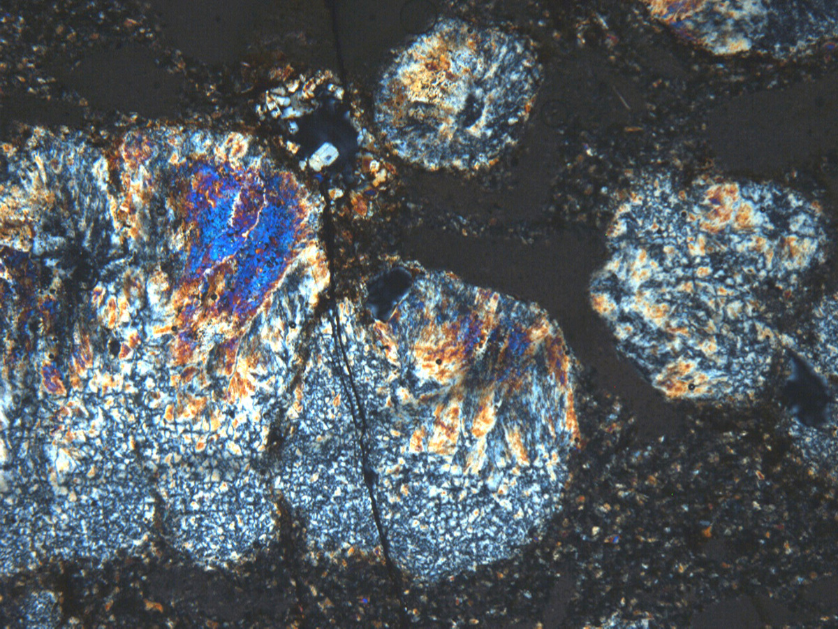
I found this thin section view (crossed polars) in my own files for chert from the McNamara Formation at McNamara locality in Missoula County, Montana. FOV is 20 mm.
Bonafide chert is at far right. No question about it (it scratches glass without even trying). In the upper middle is what resembles poikilotopic anhydrite, characterized mostly by higher first-order interference colors, up to second-order blue. Grayish material at far top is the same stuff, but where the slide is getting thinner as the edge is approached (so don’t mistake it for gypsum). At lower left of this mass is strikingly plumose material that might be also be anhydrite or maybe baryte. Other material in this thin section is siltstone containing clay chips, the latter of which only show up because of the lack of orange points of light that are quartz silt. Now, what is ultra-important here is that there are three separate globular masses of possible anhydrite. So, these are (or were) what have been called chicken-wire anhydrite nodules. They are not uncommon in evaporitic rocks, and they are known for merging together into larger, more or less continuous masses. If you look closely at the lower left of this mass, you can see where nodule formation pushed aside some dark matrix material, so these nodules formed displacively.
The next photo below shows a string of such nodules in a typical grouping. The evaporite mineral has been dominantly (or entirely) replaced by chert now, however. If you look carefully you can see some pseudo-interference crosses and squarish "windows" in the chert that has formed around this uncertain mineral. The third photo is detail of the plumose habit of this material, which I think looks a lot like some anhydrite and baryte I’ve seen in thin section. However, anhydrite tends to form clearly differentiated crystals that form in linear successions rather than in these sheafs that go on and on without any distinct breaks. Baryte tends to be more strongly colored, has greater relief, and shows botryoidal growth lines.

Strings of chalcedony after anhydrite nodules, McNamara
Plumose material grading to chert
So, chert has replaced gypsum, anhydrite, or some other evaporite-related mineral (such as possible secondary baryte), going through a stage where those squarish window-like crystallites appear.
Anybody? Come on folks give me some help here! I don’t know what I’m talking about either, so don’t be shy.
31st Oct 2018 15:53 UTCDaniel Bennett
it seems you may be in new terrain here so you may not get much help. hopefully I'm wrong. thanks for sharing. looking forward to your upcoming articles. -Daniel
3rd Nov 2018 21:33 UTCNorman King 🌟 Expert

After several years of using Mindat as a wonderful source of technical information, today I found this photo after literally days of searching for answers to some of my questions about chalcedony. This appeared in the results of an online Google search that simply duplicated an earlier one I had already done (in which it was not part of the results), except for the order of search terms. While it doesn't necessarily answer questions I had presented here, it does have more information for me on this issue in the description, and practical information in the photo itself, than any other photo on Mindat that I can remember. Granted, I had the advantage of all that preparation, or perhaps it was BECAUSE I had that preparation, that I was ready for it. I can see that it might not be as useful for beginners.
I know that the size format for POTD is not what you like, and I know that I am not a formal recommender of POTD’s, but you simply MUST make this a POTD. I’m sure you can see that you could turn it on its side and solve the first problem, adding a few minor mechanical changes to the description and turning both photos 90 degrees individually to the left (maybe Amir would agree to do the ground work), and no, you cannot simply turn the whole thing to the right--that would put the wrong colors in each quadrant. Please forget the second problem I mentioned above, since Mindat users deserve to see this! After all my work trying to find answers to various questions I have had, this single (double?) photo has taught me more than any other photo or person at Mindat.
3rd Nov 2018 23:45 UTCFrank K. Mazdab 🌟 Manager
That is a spectacular pair of photos.
For those who might be wondering if the lower photo is simply a "photo effect" (like a Snapchat filter), what it actually is is the effect of the insertion of the 1λ accessory plate (commonly called the "gypsum plate") on the regular interference 1st order gray to white interference colors (top photo) of quartz in thin section.
The extra distance the light has to travel due to the insertion of the accessory plate through the mineral results in either a constructive interference effect (where we see "blue" in the lower figure) or a destructive interference effect (where we see "orange"). The accessory plate can only be inserted in one orientation and so is a constant, thus whether this additive effect is constructive or destructive is entirely a function of the optical properties (and the orientation) of the mineral underneath.
What's interesting to me about the lower photo is that usually chalcedony fibers are enlongated along the a crystallographic axis, and due to the underlying optical properties of quartz, this mean we should see destructive interference (orange) in the NE and SW quadrants, and constructive interference (blue) in the NW and SE quadrants (assuming the photographer's accessory plate is oriented the same as mine). This orientation is termed "length fast" (because in two diagonal quadrants the vibration direction of the "fast" ray [the low refractive index] of the enlongated chalcedony is parallel to the "slow" ray vibration direction [corresponding to the high refractive index] of the material in the 1λ plate, and in the two other diagonal quadrants it's the opposite) . We do see this in the inner ring, but the colors in the outer ring are oddly reversed.
This is strange. It suggests either the fibers in the outer ring are enlongated along the c crystallographic axis (less common but certainly possible), or that somehow we're looking perpendicular to the fibers rather parallel to them. This latter scenario doesn't seem likely here. Apparently, length fast chalcedony (where we see orange in the NE) and the less common length slow chalcedony (where we see blue in the NE) can be intergrown in the same sample, so perhaps this is a wonderfully colorful example of that.
I'd vote for this to be a POTD, for its colorfulness and its educational value.
4th Nov 2018 03:01 UTCNorman King 🌟 Expert
Thanks for your vote. I don't know how the Mindat POTD process actually works. But I do know that votes from us don't make any difference. Amir Akhavan who posted this photo pair explained what is going on nicely. Here is what he said:
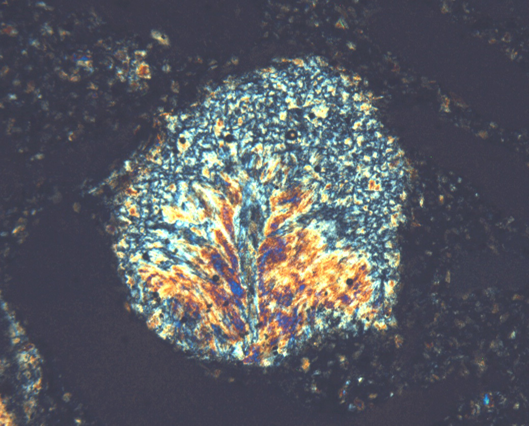
"Quartzine and length-fast chalcedony give similar extinction patterns in thin sections. Although length-fast chalcedony generally looks more fibrous and quartzine more patchy, the safe way to distinguish them is by using a λ-compensator.
Here a small spherulite of length-fast chalcedony is surrounded by a ring of quartzine, which in turn is embedded in length-fast chalcedony (note the agatoid "Runzelbänderung" in the bottom left corner).
Top: crossed polarizers
Bottom: crossed polarizers with λ-compensator.
Thin section of a gangue agate (approximate thickness 30µm).
Field of view 980µm."
Quartzine is length-slow chalcedony (other names have also been used), and it has in fact been found intergrown with length-fast chalcedony. This example is just perfect in showing the two variants and how they can be distinguished. The juxtaposition of the two types is exceptionally well illustrated here.
Much of the chalcedony I illustrated in my earlier photomicrographs is patchy, grading from only somewhat fibrous. The latter is most notable where the higher interference colors are (especially violet and blue), but also includes some grays. I still do not know how the quadrate "windows" form. I have not found references to, or illustrations of, any mineral having such crystallites. It is all fibrous, nevertheless, as the pseudo-interference crosses indicate, so we know it is chalcedony. It is length-fast. I interpret it as filling cavities formed by dissolution of gypsum/anhydrite, probably late in the diagenetic history of these rocks.
5th Nov 2018 03:34 UTCNorman King 🌟 Expert
Thanks again for sending this material to me. See what you've done? My experience is that when people slow down and look closely at something, all sorts of unexpected things show up. That's what has happened here. Mostly, people are oversubscribed and too busy to stop and look around. Glad you did!
The north-south and east-west lines I have been referring to are within the nodules rather than between them. You can't really see them in that photo because it isn't enlarged enough. You can make some out in the photo showing a single nodule of plumose material. They're really tiny, though. These nodules are distinct enough and characteristic enough for us to say that evaporitic material was indeed present here. That appears to be new information on this part of the Belt Supergroup for this area.


Mindat.org is an outreach project of the Hudson Institute of Mineralogy, a 501(c)(3) not-for-profit organization.
Copyright © mindat.org and the Hudson Institute of Mineralogy 1993-2024, except where stated. Most political location boundaries are © OpenStreetMap contributors. Mindat.org relies on the contributions of thousands of members and supporters. Founded in 2000 by Jolyon Ralph.
Privacy Policy - Terms & Conditions - Contact Us / DMCA issues - Report a bug/vulnerability Current server date and time: April 19, 2024 02:23:44
Blende Lake Iron occurrence, MacGregor Township, Thunder Bay District, Ontario, Canada