Plaka Mine No. 145, Plaka Mines, Plaka, Lavrion mining district, Lavreotiki, East Attica, Attica, Greece
© Fritz Schreiber
A beautiful spray of pharmacolite
Lenticular subparallel plagionite crystals (SXRD-analysed) epitaxially grown on the (100) face of a galena crystal. Found on a specimen that came from a fluorite-galena-sphalerite-siderite vein close to the well-known erythrite locality. Collection NHM Vienna. Photo Harald Schillhammer. This photo was originally published in: Rieck, B., ...
© Harald Schillhammer
SEM micrograph (BSE mode) showing thin chamosite platelets (bright grey) embedded in a kaolinite-grup mineral (dark grey). On the left side there is some relic pyrrhotite (bright grey). The homogeneous grey phase is calcite. Polished section of a pyrrhotite-rich ore specimen from the well-known "erythrite locality". This material was ...
© Uwe Kolitsch
Golden-brown Ranciéite formed on stalactites of several undefined Mn and Fe oxides.
© Steve Rust
Pale green to nearly white annabergite on a fuchsite-rich matrix. The ID of annabergite has been confirmed by analysis made at the Naturhistorisches Museum Wien by HR Dr. Vera Hammer.
© F.Schreiber
FOV 10mm Aug.2006
FOV 3mm
Dolomite on schist matrix with fuchsite FOV: 10mm
Tiny white balls of annabergite on a crystal of sphalerite. The ID of annabergite was established at the Museum of Natural History, Vienna. FOV: 1.5mm
This specimen is a present from my friend Vasilis
Aug.2006 small white to pale greenish balls are annabergite
Baryte cristal on Ankerite Field is 2,08 x 1,35 mm Zeiss luminar 25 mm on bellows + nikon D1x. CZM Photo & collection Chollet Pascal (# 1305)
© Chollet Pascal
Pyrite balls, consisting of crystals that evenly display cube and octahedron faces on calcite. FOV: 4mm
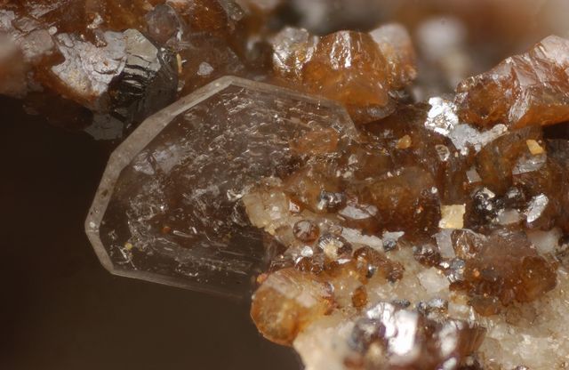
The Adit #145 has yielded some phantastic clear barytes. Nikon D70, Helicon Focus FOV: 11mm
FOV 5mm
Pink erythrite with brown resinous smolyaninovite on schist matrix.
SEM micrograph (BSE mode) of a polished section of the fuchsite-rich carbonate-schist matrix of the small white pustule-like annabergite aggregates on the roof of a large underground hall (see https://www.mindat.org/photo-244091.html). Ankerite as a grey, late-stage(?) filling of a narrow crack in slightly zoned dolomite (darker grey). Minor ...
© Michael Samouhos
iridescent pyrite on calcite.
photo and collection: hans-Jürgen Haas
Sceptre Quartz , p.w. 7.46 mm , Coll. David Nieto , Photo Ko Jansen.
© Ko Jansen
Brassy pyrite on calcite. FOV: 4mm
SEM micrograph (BSE mode) showing pentlandite (bright), with measured compositions (at.%) of Ni29.3 Fe25.5 S46 and Ni30.2 Fe24.2 S45.6, intergrown with Ni-bearing pyrrhotite (bright grey; Fe46.4 Ni1.8 S52) and pyrite (grey), in a matrix of calcite and quartz. Polished section of a pyrrhotite-rich ore specimen from the well-known "erythrite ...
SEM micrograph (BSE mode) of a polished section of the fuchsite-rich carbonate-schist matrix of the small white pustule-like annabergite aggregates on the roof of a large underground hall (see https://www.mindat.org/photo-244091.html ). Gersdorffite aggregates (grey) are embedded in slightly zoned dolomite (dark grey) intergrown with minor ...
Yellow mimetite on gossan matrix
SEM micrograph (BSE mode) showing both fresh titanite (small grey, rounded grains, bit NE of centre and in lower left) and several pseudomorphs of fine-grained TiO2 (probably rutile) (grey) and calcite (medium-grey) after titanite. The three bright grains in the lower left are, from bottom to top (see also linked micrograph), Fe-rich violarite, ...
SEM micrograph (BSE mode) of a polished section of the fuchsite-rich carbonate-schist matrix of the small white pustule-like annabergite aggregates on the roof of a large underground hall (see https://www.mindat.org/photo-244091.html). Fuchsite as micaceous aggregates (grey) forming the matrix of anhedral sphalerite grains (bright). The matrix ...
SEM micrograph (BSE mode) of a polished section of the fuchsite-rich carbonate-schist matrix of the small white pustule-like annabergite aggregates on the roof of a large underground hall (see https://www.mindat.org/photo-244091.html). Subhedral gersdorffite aggregate (grey) with tiny included galena grains (white), in a dolomite matrix. This ...
SEM micrograph (BSE mode) of a polished section of the fuchsite-rich carbonate-schist matrix of the small white pustule-like annabergite aggregates on the roof of a large underground hall (see https://www.mindat.org/photo-244091.html). Ullmannite (small white grain) next to gersdorffite aggregates (bright grey), both in slightly zoned ...
Pseudomorphose of Goethite after Pyrite now brownish/black , p.w. 2.40 mm , Coll. David Nieto , Photo Ko Jansen.
Stibnite Crystals metal grey , p.w. 3.56 mm , Coll. David Nieto , Photo Ko Jansen.
SEM micrograph (BSE mode) showing cobaltite grains (bright grey, with tiny white galena inclusions) embedded in pyrrhotite (grey) or the dark grey matrix of quartz. Polished section of a pyrrhotite-rich ore specimen from the well-known "erythrite locality". The polished section was made to identify the previously unknown cobalt source of the ...
SEM micrograph (BSE mode) of a polished section of the fuchsite-rich carbonate-schist matrix of the small white pustule-like annabergite aggregates on the roof of a large underground hall (see https://www.mindat.org/photo-244091.html). Tiny ullmannite (white) rimmed by gersdorffite (bright grey), in a dolomite-quartz matrix. This material was ...
SEM micrograph (BSE mode) showing zoned micaceous chamosite aggregates (+/- grey, with slightly variable Fe:Mg ratio) surrounding anhedral pyrite (bright grey) intergrown with some chalcopyrite (bright), in quartz. Polished section of a pyrrhotite-rich ore specimen from the well-known "erythrite locality". This material was described ...
SEM micrograph (BSE mode) showing three bright grains: from bottom to top they are: Fe-rich violarite, chalcopyrite and zircon. On the left a pseudmorph of TiO2 (probably rutile) and calcite after titanite. The matrix is quartz. Polished section of a pyrrhotite-rich ore specimen from the well-known "erythrite locality". This material was ...
Mimetite spays on Goethite "branches". No analysis. Just visual identification.
© Alexandros Frantzis
...nothing left of the fluorite that was initially covered by the dolomite. Now there is only the frame left.
© F. Schreiber
SEM micrograph (BSE mode) showing fine-grained aggregates of Ca- and Mg-bearing siderite (bright grey) intergrown with calcite (grey) and a fine-grained kaolinite-group mineral (dark grey). The brighter grey, rounded grain in the upper left is fluorapatite. Polished section of a pyrrhotite-rich ore specimen from the well-known "erythrite ...
SEM micrograph (BSE mode) showing an euhedral zircon crystal (bright) next to a fluorapatite crystal (grey). The small grey grains in the rest of the picture are TiO2 (probably rutile) in calcite (= pseudomorph after titanite). In the bottom area, micaceous chamosite (darker grey) sits in a matrix of fine-grained muscovite (dark). Polished ...
SEM micrograph (BSE mode) of large scheelite grain (white), rimmed by calcite (dark grey), associated with a small fluorapatite grain (grey, above centre) and a cobaltite grain (bright grey, lower left), in quartz. There is also some anhedral calcite (dark grey) in the lower part of the picture. Polished section of a pyrrhotite-rich ore specimen ...
photo and collection: Hans-Jürgen Haas
Valentinite on calcite.
© Heyninck A
Mimetite on cerussite
SEM micrograph (BSE mode) showing small, more or less euhedral cobaltite grains (bright grey) embedded in pyrrhotite (grey). The tiny white phase is galena. Polished section of a pyrrhotite-rich ore specimen from the well-known "erythrite locality". The polished section was made to identify the previously unknown cobalt source of the erythrite - ...
SEM micrograph (BSE mode) showing a spray of coarse-grained muscovite (grey), locally intergrown with a kaolinite-group mineral (dark). The bright grey grain is Ni-bearing pyrite. The matrix is quartz (grey). The dark phase is resin. Polished section of a pyrrhotite-rich ore specimen from the well-known "erythrite locality". This material was ...
SEM micrograph (BSE mode) showing two large fluorapatite grains (bright grey) in a matrix of predominantly calcite (grey, mainly on left) and a kaolinite-group mineral (dark, fine-grained) with inclusions of small, platy chamosite (grey). Polished section of a pyrrhotite-rich ore specimen from the well-known "erythrite locality". This ...
SEM micrograph (BSE mode) showing large agglomerates of subhedral fluorapatite grains (bright grey) in a matrix of quartz (dark grey). The fluorapatite all seem to show the same orientation, suggesting that they originally formed one large crystal that was later fractured by hydrothermal fluids. There is also minor calcite (grey) in the centre and ...
SEM micrograph (BSE mode) of a polished section of the fuchsite-rich carbonate-schist matrix of the small white pustule-like annabergite aggregates on the roof of a large underground hall (see https://www.mindat.org/photo-244091.html). Small millerite grain (grey) surrounded by ullmannite (bright grey), next to gersdorffite (medium grey), all in ...
Processed by: Helicon Filter;
Stibnite Crystals metal grey , p.w. 0.94 mm , Coll. David Nieto , Photo Ko Jansen.
Clear baryte crystals with little pyrite. This specimen is a present from a good Greek friend, Vasilis.
SEM micrograph (BSE mode) of a polished section of the fuchsite-rich carbonate-schist matrix of the small white pustule-like annabergite aggregates on the roof of a large underground hall (see https://www.mindat.org/photo-244091.html). Minor small ullmannite (white) and major gersdorffite (bright grey) rim an Fe-bearing, distinctly zoned ...
Stibnite Crystals metal grey ,supported by a Calcite crystal white and an other in right corner , p.w. 1.42 mm , Coll.David Nieto , Photo Ko Jansen.
Calcite Crystals ,clear/transparent , p.w. 6.17 mm ,Coll. David Nieto , Photo Ko Jansen.