Home PageAbout MindatThe Mindat ManualHistory of MindatCopyright StatusWho We AreContact UsAdvertise on Mindat
Donate to MindatCorporate SponsorshipSponsor a PageSponsored PagesMindat AdvertisersAdvertise on Mindat
Learning CenterWhat is a mineral?The most common minerals on earthInformation for EducatorsMindat ArticlesThe ElementsThe Rock H. Currier Digital LibraryGeologic Time
Minerals by PropertiesMinerals by ChemistryAdvanced Locality SearchRandom MineralRandom LocalitySearch by minIDLocalities Near MeSearch ArticlesSearch GlossaryMore Search Options
The Mindat ManualAdd a New PhotoRate PhotosLocality Edit ReportCoordinate Completion ReportAdd Glossary Item
Mining CompaniesStatisticsUsersMineral MuseumsClubs & OrganizationsMineral Shows & EventsThe Mindat DirectoryDevice SettingsThe Mineral Quiz
Photo SearchPhoto GalleriesSearch by ColorNew Photos TodayNew Photos YesterdayMembers' Photo GalleriesPast Photo of the Day GalleryPhotography
╳Discussions
💬 Home🔎 Search📅 LatestGroups
EducationOpen discussion area.Fakes & FraudsOpen discussion area.Field CollectingOpen discussion area.FossilsOpen discussion area.Gems and GemologyOpen discussion area.GeneralOpen discussion area.How to ContributeOpen discussion area.Identity HelpOpen discussion area.Improving Mindat.orgOpen discussion area.LocalitiesOpen discussion area.Lost and Stolen SpecimensOpen discussion area.MarketplaceOpen discussion area.MeteoritesOpen discussion area.Mindat ProductsOpen discussion area.Mineral ExchangesOpen discussion area.Mineral PhotographyOpen discussion area.Mineral ShowsOpen discussion area.Mineralogical ClassificationOpen discussion area.Mineralogy CourseOpen discussion area.MineralsOpen discussion area.Minerals and MuseumsOpen discussion area.PhotosOpen discussion area.Techniques for CollectorsOpen discussion area.The Rock H. Currier Digital LibraryOpen discussion area.UV MineralsOpen discussion area.Recent Images in Discussions


Mineral ExchangesWill trade specimen for photomicrography of it

8th Dec 2014 20:14 UTCOlivier Langelier

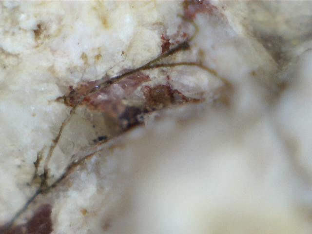
I have specimen that way too beautiful for my cheap digital microscope. One example
is very interesting vermicular Spinel.
As the title says, I will trade such specimen in return for good photomicrograph of it.
If you're interested simply send me a private message with your mail address as well
as a couple photomicrograph to show what your equipment can do.
Thanks!


8th Dec 2014 21:09 UTCReiner Mielke Expert
8th Dec 2014 22:38 UTCOlivier Langelier

Here's another example, different color same structure

9th Dec 2014 00:15 UTCReiner Mielke Expert
9th Dec 2014 01:09 UTCOlivier Langelier

on these images is around 1mm
There's just too many occurrences on too many specimen coming from several
sources for it to be a coincidence.
9th Dec 2014 14:26 UTCReiner Mielke Expert
9th Dec 2014 17:09 UTCOlivier Langelier
look it up? http://www.alexstrekeisen.it/english/vulc/olivinexenocryst.php has nice
examples. The macle chain structure is very visible on many of my pictures.
I'm ressourceful, I'll find a way to get good enough pictures to prove it, even if no one
here helps.
9th Dec 2014 17:37 UTCReiner Mielke Expert

9th Dec 2014 18:31 UTCNorman King 🌟 Expert
You show fibres in the air (so far I am still not at the point of denying they could be mineral fibres). But the illustrations in the article are fibres in solid mineral matter. Those would not likely weather out in 3-D relief, as what you show. Moreover, they are tightly packed, with multiple examples together. Lone fibres in air are not the same thing.
As an independent observation, you show what looks just like fibres from textiles or perhaps paper or insulation. I find the same sort of thing on most specimens I photograph, and I can manipulate them to clean them off before photography if I can do so without damaging delicate nearby minerals. And, if necessary, I use Photoshop to get rid of them in the photos because they are so distracting. So, now, using good judgment, I should say they are not mineral fibres.
We have to evaluate: Which do you think is more likely–individual loose fibres of materials adhering to mineral specimens that have spent time in soil, boxes, papers, or other wrapping material, or an obscure and exotic texture of tightly packed fibres around olivine grains within particular igneous rocks that have been displayed for us only in petrographic thin sections?
9th Dec 2014 20:22 UTCbill wall
10th Dec 2014 00:26 UTCOlivier Langelier

Even on my crappy pictures it's so visible


10th Dec 2014 14:17 UTCReiner Mielke Expert
10th Dec 2014 16:52 UTCDoug Daniels
1) How do you know the things are spinel - have they been analyzed?
2) The photos shown by Strekeisen are of thin sections, taken through a petrographic microscope, thus show different things than you will see in a "raw" specimen. Also, his "vermicular spinel" occurs with an orthopyroxene - is that also present in your samples?
3) And, if the specimens are of mantle peridotite, where were they collected from? Can give credence to your arguments.
10th Dec 2014 16:53 UTCOlivier Langelier

have a reading and a SEM image of this thing. For the rest it's Olivine and serpentinization
products, Pyrope, Spinel, Chromite
I tell you, it's not just fibers, sooner or later I'll find a way to get a picture good enough to
prove it conclusively. I also used the latest photo software and compared the blue color of
the known spinel to that of the presumed spinel filaments and oddities and it's a perfect match
I agree that the pictures on the website I have linked are not the best but that's the only
I could find to compare.

10th Dec 2014 17:26 UTCReiner Mielke Expert
10th Dec 2014 18:26 UTCOlivier Langelier
What I want is to see it better! Any reasonnable photomicrography setup will do
a better job than my ridiculous digital microscope. I don't care for a thin section
I want to see the 3D structure of it in color
10th Dec 2014 20:25 UTCRalph S Bottrill 🌟 Manager


Mindat.org is an outreach project of the Hudson Institute of Mineralogy, a 501(c)(3) not-for-profit organization.
Copyright © mindat.org and the Hudson Institute of Mineralogy 1993-2024, except where stated. Most political location boundaries are © OpenStreetMap contributors. Mindat.org relies on the contributions of thousands of members and supporters. Founded in 2000 by Jolyon Ralph.
Privacy Policy - Terms & Conditions - Contact Us / DMCA issues - Report a bug/vulnerability Current server date and time: April 26, 2024 18:45:00