Julian Gray's Photo Gallery
CDQ-7AUOregonite Ni2FeAs2

Dimensions: 20 mm x 11 mm x 4 mm
NK0-XD1Malachite Cu2(CO3)(OH)2
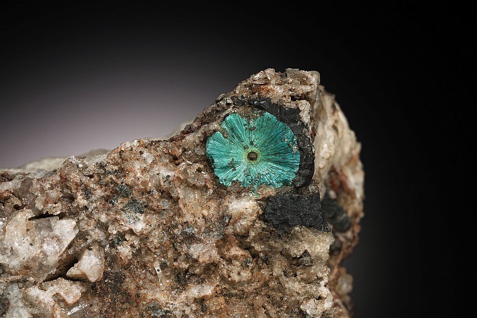
Largest Crystal Size: 1.0 cm
Specimen from the Tellus Science Museum Collection (TL2009.22.2127) and the former Geology Collection of the Georgia Capitol Museum. This specimen was depicted on page 185 of the Minerals of Georgia (Cook and Gray, 2016)
Y6Y-92TRutile TiO2

Dimensions: 4 cm x 3.3 cm x 2.3 cm
From the Tellus Science Museum collection (MIN-3433). Photograph depicted on page 247 of Minerals of Georgia (Cook, R.B., and Gray, J.C. 2016).
7UW-LT5Vauxite Fe2+Al2(PO4)2(OH)2·6H2O

Dimensions: 1 mm x 1 mm x 1 mm
T4T-A6PVauxite Fe2+Al2(PO4)2(OH)2·6H2O

Dimensions: 1 mm x 1 mm x 1 mm
Modified by CombineZP
2XT-XRPVariscite AlPO4·2H2O

Transparent variscite crystal approximately 2 mm wide. The variscite is in a cavitiy of a leached lazulite crystal. Matrix is quartz sericite with finely dissiminated rutile and pyrite. A 0.25 mm pyrite crystal (cube and pyritohedron habit)is visible in the right foreground.
Specimen collected by Ed and Martha Cunnignham. Photographed using a Canon T3i SLR mounted on a bellows and a 4 x microscope objective. The image is a composite of 31 stacked images taken with 50 micron separation between each stack. Final image processed using Combine ZP.
CKR-9GGVariscite AlPO4·2H2O

Transparent variscite crystals approximately 2.5 mm wide. The small red inclusions are rutile. The variscite is in a cavitiy of a leached lazulite crystal. Matrix is quartz sericite with finely dissiminated rutile and pyrite.
Specimen collected by Ed and Martha Cunnignham. Photographed using a Canon T3i SLR mounted on a bellows and a 4 x microscope objective. The image is a composite of 15 stacked images taken with 50 micron separation between each stack. Final image processed using Combine ZP.


